Verdich 7 1 year ago. Which image represents cytokinesis in an animal cell.

Mitosis Flashcards Quizlet
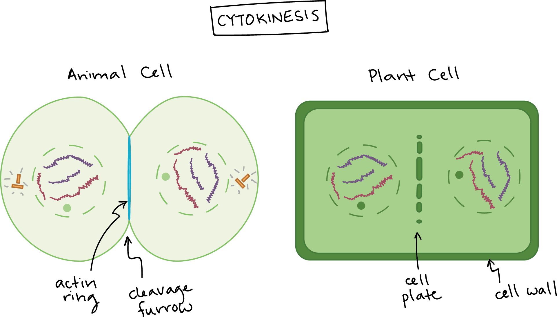
Cytokinesis the final step in cell division partitions the contents of a single cell into two.
. Scanning electron micrograph of just-divided hela cells. Which image represents cytokinesis in an animal cell. You might be interested in.
Science - 4th. Which image represents cytokinesis in an animal cell. Which organelle labeled x in the diagram is found in both plant and animal cells.
Cytokinesis in animal cells begins during anaphase as a cleavage furrow an indentation of the membrane. Which part of the cell cycle does this image represents in an animal cell. Microtubules are depicted in blue the actomyosin contractile ring and the midbody ring in red and the endosomal sorting complex required for transport ESCRT-III spiral filaments in green.
1 MULTIPLE CHOICE OPTION. Which image represents cytokinesis in a plant cell. In most cells cytokinesis is initiated during the.
Lasso-like filaments constrict to deepen the furrow until the cytoplasm is separated between the two daughter cells. Spindle fibres microtubules and chromosomes are still visible. Mitosis and Cytokinesis in Animal Cells.
Cytokinesis the final step in cell division partitions the contents of a single cell into two. In à animal cells the plasma membrane of the cell parent pinch inward along the cell of the cell until two daughters are formed. In animal cells cytokinesis is accomplished by furrow formation which starts from the cell membrane.
Arthur made a mistake labeling the diagram of the three stages of the cell cycle. A new cell membrane is generated from the vesicles of Golgi. A teacher makes a venn diagram.
Only one image is a plant cell. Answerthe third image represents. Cytokines is the third image.
Plant cells undergo cytokinesis by forming a new cell wall between the daughter cells. Mitosis has five stages that are usually associated with it. - cytokinesis stock pictures.
Cytokinesis is the final process in eukaryotic cell division which divides the cytoplasm organelles and cellular membraneCytokinesis typically occurs at the end of mitosis after telophase but the two are independent processesIn most animals cytokinesis begins sometime in late anaphase or early telophase to ensure the chromosomes. Jacqueline trying to draw an image of a cell in telophase. - cytokinesis stock pictures royalty-free photos images.
Which describes what she should draw. Meanwhile Cytokinesis in animal cells produces new cell membranes from the Endoplasmic reticulum. Cytokinesis in Plant Cells.
This step even though it is not directly related to mitosis is important for mitosis to begin. Cytokinesis in Animal Cells. Prisoha 69 1 year ago.
I promise you I took the test and thats an animal cell not olant So its D. It is not c. Which image represents cytokinesis in a plant cell.
In this process cleavage of cytoplasm takes place. NOT a cell with chromosomes pulling apart. An animal cell left and a plant cell right are shown.
When it comes to the spindle the spindle middle part stays active and vigorous during cytokinesis in plant cells but in animal cells the Cytokinesis spindle degenerates. Cytokinesis relies on a tight interplay between signaling and cellular mechanics and has attracted the attention of both biologists and physicists for more than a century. Schematic diagram illustrating the different stages of cytokinesis in animal cells.
Figure 41313 Mytotic cytokinesis. Plants and Animal Adaptations. Examine the images of a plant cell in the different stages of mitosis.
In animals cytokinesis is centripetal. Which describes what she should draw. Which step of mitosis involves the condesing of DNA into chromosomes.
Jacqueline is trying to draw an image of a cell in telophase. Which image represents cytokinesis in a plant cell. Cytokinesis relies on a tight interplay between signaling and cellular mechanics and has attracted the attention of both biologists and physicists for more than a century.
Cytokinesis is a physical process of cell division that normally takes place after mitosisCytokinesis is the physical division of the cell cytoplasm the cell membrane and cell organelles in eukaryotic cells to produce two distinct cells at the end of the cell cycle in both mitosis and meiosis. In animal cells cytokinesis occurs through cortical remodeling orchestrated by the anaphase spindle. During the cytokinesis the cytoplasm is divided into two and the cell is divided as shown below.
Which are the main stages of the cell cycle. Which image represents the step in mitosis when chromosomes condense and spindle fibers form. Whitefish mitosis whitefish embryo blastula telophase cytokinesis daughter cells magnification x250 cleavage furrow has constricted the cell into two daughter cells.
Report an issue. The cytokinesis is the final phase of the cell division. In animal cells cytokinesis occurs through cortical remodeling orchestrated by the anaphase spindle.
Which image represents cytokinesis in an animal cell. Thus in animal cells cytokinesis starts from. The first is called interphase in which an animal cell prepares for reproduction by maturing and replicating its chromosomes.
3 MULTIPLE CHOICE OPTIONS. Whats the answer omg hint plant cells are always green its d the square shaped cell wall indicates that it is a plant cell. The 3rd image good luck Send.

Which Image Represents Cytokinesis In An Animal Cell The 2 I M Choosing Between I Saw Mixed Brainly Com

File Plant And Animal Cell Cytokinesis Svg Wikipedia
Phases Of Mitosis Mitosis Biology Article Khan Academy

Which Image Represents Cytokinesis In An Animal Cell Brainly In

Mitosis Ck 12 Foundation

Which Image Represents Cytokinesis In An Animal Cell Brainly In

1 Which Letter Represents Mitosis And Cytokinesis Ppt Download

Which Image Represents Cytokinesis In An Animal Cell Brainly In
0 comments
Post a Comment